Views: 0 Author: Site Editor Publish Time: 2026-07-13 Origin: Site


Indirect ophthalmoscopes and direct ophthalmoscopes are two common ophthalmic examination tools, differing significantly in several aspects. The following is a detailed comparison of these two types of ophthalmoscopes:
I. Structure and Principle
Indirect Ophthalmoscope:
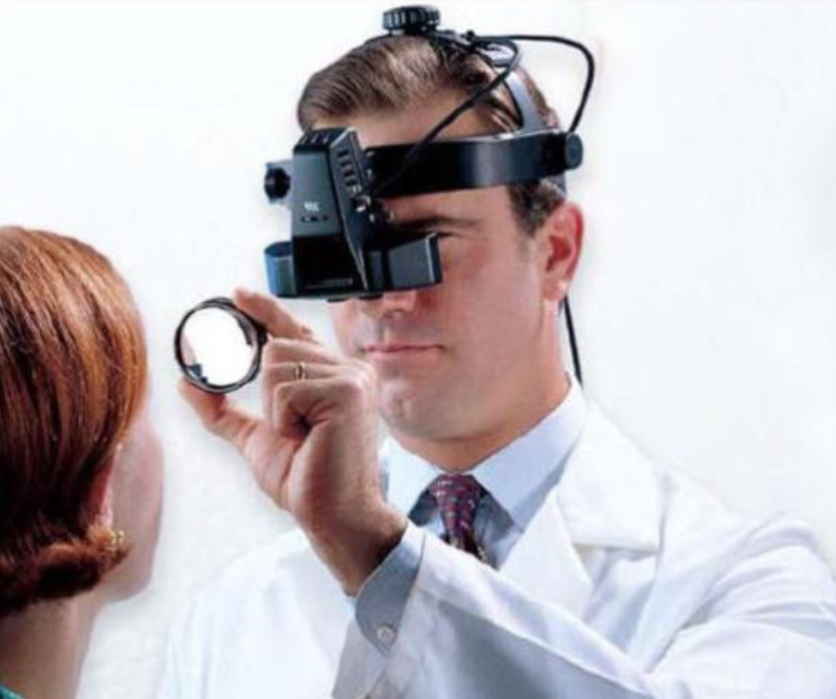
Structure: The main part of an indirect ophthalmoscope consists of a specially designed light source and eyepiece fixed to a plastic headband. A +20D convex lens is typically used to magnify the fundus before examination.
Principle: Light enters the eye being examined through the pupil, and the reflected light forms an image in front of the patient's eyes using a focusing lens.
Direct Ophthalmoscope:
Structure: A direct ophthalmoscope consists of a handle, lens, and power supply, including an observation system and an illumination system. The illumination system includes a focusing lens, a bulb, and a reflecting mirror. The observation system includes a focusing system and a peephole.
Principle: Light enters the eye being examined through the pupil, and the reflected light forms an image inside the patient's eye.
II. Examination Methods and Characteristics
Indirect Ophthalmoscopy:
Examination Method: During the examination, the examiner should maintain a certain distance from the patient (e.g., an arm's length) and use a binocular indirect ophthalmoscopy.
Characteristics:
Relatively low magnification, but a wide field of view, facilitating comprehensive observation of the fundus.
The image is an inverted real image with a stereoscopic effect.
High illumination brightness, suitable for patients with poor transparency of the refractive media.
Pupillary dilation is required during the examination.
Direct Ophthalmoscopy:
Examination Method: During the examination, the examiner's eyes must be close to the patient's eyes. The examiner's other hand pulls back the patient's eyelids, and the ophthalmoscopy is placed at a certain distance in front of the patient's eyes for examination.
Features:
High magnification, facilitating observation of fine fundus structures.
Image is an upright virtual image.
Relatively low illumination, unsuitable for patients with opacities in the refractive media.
Mydriasis is usually not required during examination.
III. Applications and Advantages
Indirect Ophthalmoscope:
Applications: Primarily used for examining retinal detachment, fundus tumors, and pediatric fundus examinations.
Advantages: Wide field of view, high illumination, strong stereoscopic effect, facilitating the detection of retinal lesions.
Direct Ophthalmoscope:
Applications: Primarily used to examine the posterior pole of the fundus, such as the optic disc, macula, and retinal vessels.
Advantages: Simple operation, high magnification, and portable.