


| Quantity: | |
|---|---|


LBW-186 EEG monitor
Using the aEEG (amplitude-integrated EEG)+EEG (original EEG) to integrate the blood oxygen monitoring and video monitoring functions.Help the clinicians understand the brain health of children in time,provide a diagnosis basis for children with brain injury to receive early intervention,and reduce or avoid secondary brain injury.
CLINICAL APPLICATION OF BRAIN FUNCTION MONITORING
SCREENING
Screening neonates at risk of neonatal hypoxic ischemic encephalopathy(HIE), septicemia of preterm infants (SIE), asphyxia,epilepsy,convulsions,intracranial hemorrhage and hydrocephalus.
MONITORING
Real-time monitoring of the dynamic changes in the neonatal brain function,and early detection of corresponding change trends;
Sleep-wake cycle monitoring in term and preterm infants;
Timely detection of subclinical convulsion, epileptic seizure and epileptic state;
Monitoring the course and efficacy of hypothermia therapy in HIE.
ASSESSMENT
Assess the therapeutic effects of various interventions;
Assess the degree of brain damage in patient and improve the accuracy of prognosis;
Assess the brain maturity in patient;
Timely conduct clinical intervention in the reversible stage of brain function damage;
Assist in the assessment of brain death.
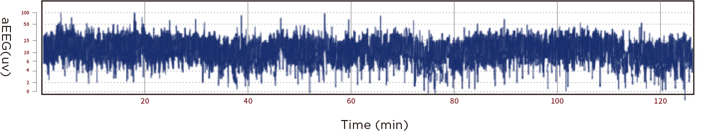
Amplitude-integrated ECG (aEEG)
⦁ Intuitive and easily interpreted spectrum
aEEG is a simplified form of continuous recording of EEG.It is to express the change signal of the original brain wave amplitude in semi-logarithmic form after filtering,amplitude integration and time compression,and to reflect the overall level of the background activity of the cerebral cortex
through the upper and lower boundaries of the spectral band.
The upper boundary,lower boundary and bandwidth are three important indicators for analyzing aEEG, and 2h is taken as the optimal duration for spectrum analysis
⦁ Quickly and easily realize brain injury screening, brain function monitoring and brain development assessment in patient,providing effective auxiliary diagnosis reference for clinical treatment;
Automatically identify low voltage and provide tips to help the clinicians adjust the therapeutic regimens in time.
⦁ aEEG background activity pattern identification and classification
According to the amplitude and continuity of the upper/lower boundary of aEEG, the neonatal EEG activity will be classified,and the Hellstrom-Westas background five-classification method will be used to improve the ability of medical staff to identify the aEEG waveforms.
HIE multi-dimensional identification and application
Neonatal ischemic encephalopathy (HIE) is one of the common neurological diseases in the perinatal period and one of the diseases with a high disability rate in the neonatal period.
⦁ aEEG has high sensitivity and specificity for the early diagnosis of HIE patients and the evaluation of the degree of injury (the time window for optimal intervention is within 6 hours of the occurrence of HIE).
Take the neuroprotective measures in time to reduce the mortality and morbidity and improve the accuracy of prognosis.
⦁ Rainbow SET pulse oximetry monitoring
Monitor the SpO2(blood oxygen saturation)/PI(blood perfusion index/PR (pulse)change in a non-invasive and continuous manner,effectively eliminate the exercise interference,improve the measurement performance under low perfusion and body motion,and assist in evaluating neonatal hypoxia to guide the clinical treatment.
⦁ The upgradeable hemoglobin parameter
Perfusion variation index(PVI)enables the clinicians to assess the patient's infusion status, and measure the hemiglobin SpMet in the blood in a non-invasive and continuous manner to assess the blood oxygen content SpOC.
⦁ Burst-suppression,a severe abnormal EEG phenomenon characterized by burst of high-amplitude activity alternating with low voltage or electrical suppression.It can assist the clinicians in measuring bursts per minute (BPM) and interburst interval (IBl)to analyze the brain development of premature infants.

Results shown were calculated by combining the sensitivity and specificity of motion generated by machine and volunteer.

SpO2 Pulse oximetry module accessories
Epilepsy rapid identification and application
aEEG + Envelope Trend Chart (ENVELOPE) + TP + Video
⦁ Envelope trend chart (ENVELOPE) is used to represent the change in the EEG amplitude, which can better eliminate the artifacts caused by interference and identify epileptic and continuous manner, e ectively eliminate the exercise interference, improve the measurement performance under low perfusion and body motion, and assist in evaluating neonatal hypoxia to guide the clinical treatment. seizure events more accurately.
The envelope trend is considered a reliable method for detecting epileptic seizures. Based on half-wave analysis, it highlights the rhythmic events and filters the transient events. Quickly distinguish epileptic seizures, burst suppression, and comprehensively view the entire process of epileptic seizures for neonatal and child monitoring.
⦁ TP (total power), also known as total energy, reflects the change in the EEG amplitude, and has high sensitivity and low specificity for clinically detecting epileptic seizures.


Convulsion identification and application
EEG + burst-suppression ratio + aEEG + video
Neonatal seizures are often clinically atypical,mostly subclinical and minimal,making it extremely challenging to accurately identify and manage this problem in a busy NICU setting.Patients with convulsions have a very poor prognosis, with a 20%mortality rate in the neonatal period.
⦁ Conventional EEG combined with video monitoring is the gold standard for bedside monitoring.
⦁ Through the rapid identification of suspicious convulsive activity in the aEEG background activity pattern it can improve the ability of medical staff to identify the aEEG convulsive activity pattern,leaving the convulsion identification simpler and faster.
⦁ Single-channel or dual-channel aEEG combined with simultaneous EEG interpretation can increase the detection rate of convulsions.
Sensitivity to identify the number of seizures | Sensitivity to identify the children with seizures | |
Dual-lead aEEG plus raw EEG | 81.2% | 89.7% |
Single seizure | Recurrent seizure | Convulsive status | |
Single-channel aEEG + raw EEG/video | 76.9% | 76.5% | 91.3% |
Dual-channel aEEG + raw EEG/video | 88.5% | 83.5% | 95.7% |
Four-channel aEEG + raw EEG/video | 92.3% | 85.9% | 95.7% |
TECHNOLOGICAL UPGRADING TO HELP
THE CLINICIANS QUICKLY AND ACCURATELY INTERPRET
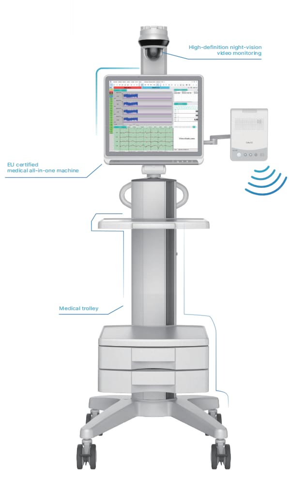
⦁ Medical all-in-one machine -"intuitive display in the multi-trend chart"
The medical all-in-one machine that complies with the EU CE-related:
EN60601-1-2:2015 and EN60601-1:2006 standards is used, with better hardware security,stability,durability and anti-interference, and the product industrial design is more in line with clinical Medical scene.
Equipped with a 19-inch color touch medical-grade display screen, displays each trend chart and corresponding values clearly and intuitively. Provides a variety of trend chart displays and real-time EEG displays to help the clinicians interpret data in time and ensure the accuracy of clinical diagnosis.
Clear event marking:Medical staff can quickly mark the events in the collection or playback process, and distinguish different events by color to effectively track the management of treatment, making the clinical evaluation more efficient,and facilitating mutual cooperation.


Equipped with rechargeable lithium-ion battery to support 2h independent working time, which is convenient for transfer of patient.
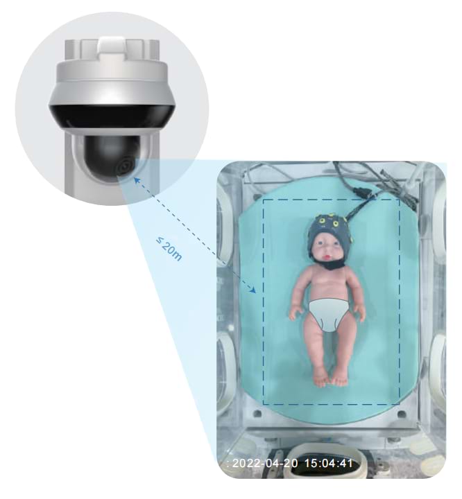
⦁ High-definition night-vision video monitoring
The optional high-definition camera with night vision function supports horizontal and vertical rotation,and the illumination distance can be up to 20m.
The physical movement of patients can be monitored in a real-time manner through the LCD screen,eliminate artifacts caused by various interference,and detects EEG signals accurately,helping the medical staff interpret the abnormal EEG activities of patients in time.
⦁ Signal amplifier -"multi-channel signal acquisition"
Standard 4 channels and optional 8/12 channels,with impedance selection button,built-in Spo2 input channel,less interference from the environment,and more stable signal acquisition.
With strong anti-interference performance,it can also record high-quality electrophysiological signals in the complex NICU and operating room environments and during the transfer process.
Provide the function of real-time detection of electrode impedance,monitor the impedance value in a real-time manner during the EEG data acquisition process,and provide the position of abnormal electrode impedance or electrode falling off in the form of electrode impedance status icon to ensure the validity of the detection information,facilitating the troubleshooting and quality control.(The EEG electrodes are placed according to the international 10-20 electrode placement standard).


The EEG electrodes are placed according to the international 10-20 electrode placement standard.
Connect with the medical all-in-one machine wired or wireless.
When the wired connection is disconnected,it will automatically switch to wireless communication mode.
The amplifier has a built-in rechargeable lithium-ion battery,
which can last for 45 min when power outage.
Power-on self-test function and a variety of fault sound-light alarm prompts.
⦁ Scientific and effective data management
Supports the EEG data playback,and supports the information management and EEG data management of patients,data transmission and storage functions,convenient for scientific research and teaching.
Automatically generate the aEEG clinical report and support the printing function,various of charts can be manually selected.


