Views: 0 Author: Site Editor Publish Time: 2026-09-28 Origin: Site


What is fundus photography?
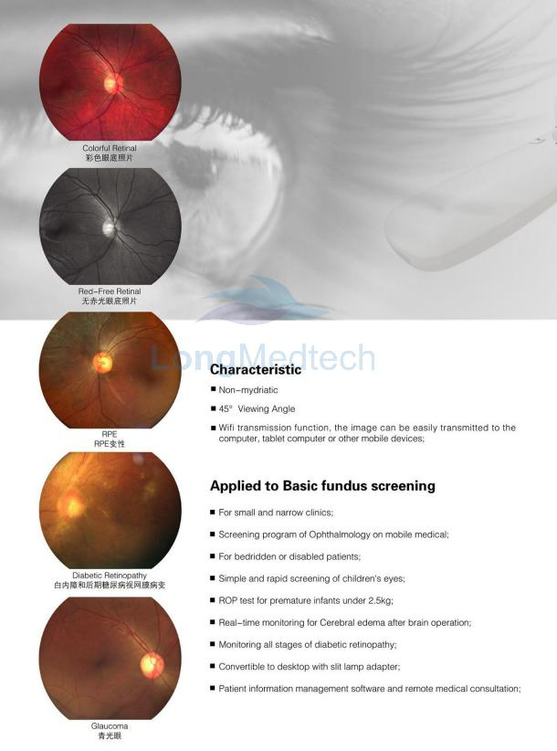
Fundus photography is the process of digitally imaging the inner surface of the eye (including the retina, macula, blood vessels and optic nerve head). It allows for the permanent preservation of an image record of the appearance of the retina.
Technology
The fundus camera uses special optical components and light to capture high-resolution images through the pupil. To improve image quality, pupil dilation may be required, but some systems can take pictures without pupil dilation for specific patients.
Depending on the equipment, the images can be in color, grayscale, or wide-angle.
Non-contact technology is usually fast and comfortable.
By comparing images from different periods, subtle changes can be detected.
It is often used in combination with other examinations (such as optical coherence tomography or angiography).
Clinical application
Fundus photography is widely used in the screening and monitoring of diabetic retinopathy, macular degeneration, glaucoma, and many other retinal diseases. It is helpful for diagnosis, patient education, and communication among healthcare professionals.
Support for remote scoring during the screening process
Helpful for documenting responses to treatments such as laser or injections
Digital storage enables side-by-side comparisons during subsequent visits.
This is crucial for the medical legal record of retinal examination results.