
Views: 0 Author: Site Editor Publish Time: 2026-05-15 Origin: Site


Intraoperative imaging in dentistry is a very specific need, meaning you can't just buy any machine that takes dental X-rays; you must choose equipment that can be used in the operating room.
Based on current clinical practice and the latest purchasing trends, the most suitable intraoperative imaging equipment for dental surgeries (such as implantology and maxillofacial surgery) usually refers to a mobile C-arm X-ray machine, rather than a traditional intraoral X-ray machine.
1. Primary Recommendation: Mobile C-arm X-ray Machine (for complex surgeries)
If your surgery involves implant placement, jaw fracture removal, cyst excision, or impacted tooth extraction, requiring real-time, dynamic, and wide-range fluoroscopic guidance, then a C-arm is standard.
Core Use: Real-time intraoperative fluoroscopy. During surgery, the dentist can step on the foot pedal at any time to see the position of instruments in the bone in real time on the monitor, such as the depth and angle of implant placement, or whether there are any bone fragments remaining.
Key Selection Criteria:
High-definition large flat panel: This is the primary requirement. A large-format (approximately 30cm x 30cm) high-definition silicon flat panel can cover a large surgical field in one operation, avoiding repeated fluoroscopy during surgery. Unclear images can easily lead to surgical errors.
Low Radiation: Multiple short exposures are required during surgery; low radiation not only protects both doctors and patients but is also a sign of advanced equipment technology.
Easy Operation: Equipped with a 4K high-definition monitor, hand-cranked and foot-operated switches, and laser positioning, facilitating operation in a sterile area during surgery.
Applicable Scenarios: Dental implantology (especially full/partial mouth implants), maxillofacial surgery, temporomandibular joint surgery.
2. Auxiliary Equipment: Digital Intraoral X-ray Machine (for pre-/post-operative confirmation) For routine intraoperative confirmation of root canal treatments, or simple pre- and post-operative verification of single tooth extractions/implants, wall-mounted or upright intraoral X-ray machines are basic and essential equipment.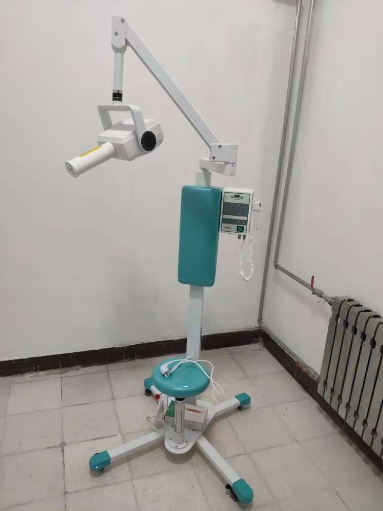
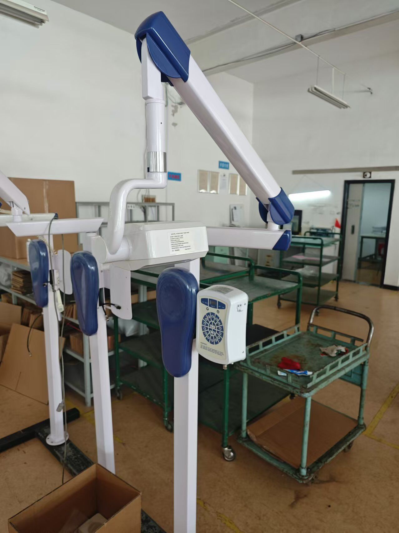
Core Use: Static, localized, high-resolution imaging. Used to take small radiographs (periapical radiographs) of single or several teeth, with extremely high resolution, clearly showing root morphology, number of root canals, and alveolar bone details. Key Selection Criteria:
Digital Readiness: Must be compatible with digital radiography (DR) sensors, enabling instant image capture and processing without the need for film processing. This is crucial for improving surgical efficiency.
High-Frequency Constant Voltage Generator: Higher frequencies (e.g., ≥150kHz) result in more stable X-rays, better image quality, and lower patient radiation dose.
Precise Parameter Adjustment: Must be able to select different exposure times for adults children and for anterior teeth/molars, avoiding images that are too bright or too dark due to differences in bone density.
Flexible Robotic Arm: At least 3 degrees of freedom in the joints ensure easy positioning next to the operating chair and prevent drifting after locking. This is crucial for smooth intraoperative positioning.
Applicable Scenarios: Root canal treatment (intraoperative confirmation of working length), post-extraction examination, and post-operative confirmation of single implant placement.
3. Equipment Not Required: Extraoral Imaging Equipment (CBCT/Panoramic X-ray Machine)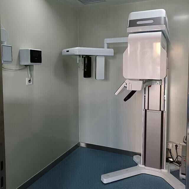
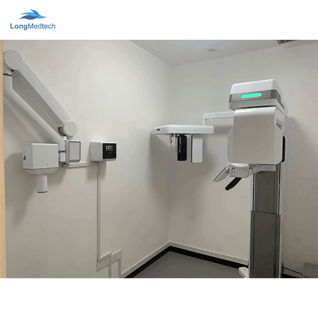
Reason: While these devices (including cone-beam computed tomography (CBCT) and panoramic X-ray machines) have high diagnostic value, they are large, require fixed installation, and cannot be brought into the operating room for real-time intraoperative guidance. They are typically used for preoperative planning and postoperative follow-up, not for "intraoperative imaging."


